Special Tests for Cervical Neck Pain: A Comprehensive Guide.
- PHYSIO 360

- Apr 11, 2025
- 3 min read

INTRODUCTION;
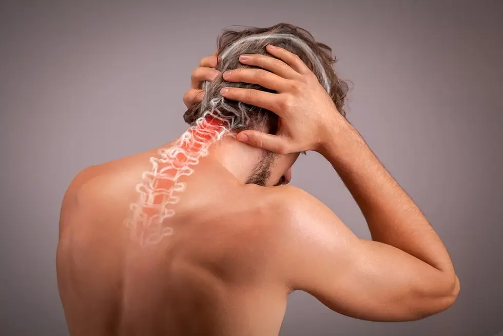
Cervical neck pain is a common complaint that affects people of all ages, whether due to poor posture, muscle strain, trauma, degenerative changes, or nerve compression. Accurate diagnosis is crucial for effective treatment, and while clinical symptoms guide the initial assessment, special tests play a vital role in confirming suspicions and narrowing down the cause of cervical dysfunction.
This blog explores the most commonly used special tests for cervical neck pain, their purpose, how they’re performed, and what positive results indicate.
Why Special Tests Matter in Cervical Pain Diagnosis.
Special tests are clinical examination techniques designed to isolate specific structures, like nerve roots, intervertebral discs, or facet joints, and determine their involvement in the patient’s pain. These tests help differentiate between:
Muscular issues
Joint dysfunction
Radiculopathy (nerve root involvement)
Ligamentous injury
Vertebrobasilar insufficiency

1. Spurling’s Test (For Cervical Radiculopathy).
Purpose:
To detect cervical nerve root compression or irritation.
How it's performed:
The patient is seated. The examiner extends the neck, rotates it to the affected side, and applies gentle axial compression.
Positive Sign:
Radiating pain or numbness down the arm on the same side.
2. Distraction Test.
Purpose:
To relieve pressure on cervical nerve roots.
How it's performed:
With the patient seated, the examiner gently lifts the patient’s head vertically (traction force).
Positive Sign:
Reduction or relief of radiating symptoms suggests nerve root compression.
3. Jackson’s Compression Test.
Purpose:
To identify nerve root impingement.
How it's performed:
The patient rotates their head to one side. The therapist applies a downward axial load.
Positive Sign:
Radicular pain on the side of the head indicates nerve root compression.
4. Shoulder Abduction Test.
Purpose:
To assess nerve root irritation, especially at C4–C6 levels.
How it's performed:
The patient is asked to abduct their arm and rest the hand on top of their head.
Positive Sign:
Reduction in symptoms (especially pain or tingling in the arm) indicates nerve root compression relief.
5. Lhermitte’s Sign
Purpose:
To detect spinal cord pathology or cervical myelopathy.
How it's performed:
With the patient seated, the examiner passively flexes the cervical spine.
Positive Sign:
An electric shock-like sensation down the spine and into the limbs suggests upper motor neuron involvement or multiple sclerosis.
6. Vertebral Artery Test
Purpose:
To assess the patency of the vertebral arteries and screen for vertebrobasilar insufficiency.
How it's performed:
The patient is positioned supine. The examiner extends, rotates, and laterally flexes the head, holding it for 30 seconds while monitoring for symptoms.
Positive Sign:
Dizziness, nystagmus, nausea, visual disturbances, or loss of consciousness—the test must be performed with caution.
7. Alar Ligament Stress Test
Purpose:
To assess the integrity of the alar ligament.
How it's performed:
The patient is seated. The examiner stabilizes C2 and side-bends or rotates the head.
Positive Sign:
Excessive movement or delay in movement of C2 suggests ligament laxity or rupture.
8. Sharp-Purser Test
Purpose:
To assess for atlantoaxial instability, especially in cases like rheumatoid arthritis.
How it's performed:
The patient is seated. The examiner stabilizes C2 with one hand and gently applies posterior force on the forehead.
Positive Sign:
A clunk or reduction in symptoms may indicate subluxation of the atlas on the axis. This test should be performed with extreme care.
9. Adson’s Test
Purpose:
To detect thoracic outlet syndrome affecting neurovascular structures in the cervical region.
How it's performed:
The patient turns their head toward the test arm, extends the neck, and inhales deeply while the examiner palpates the radial pulse.
Positive Sign:
Diminished or absent radial pulse, or reproduction of symptoms (numbness, tingling, heaviness).

When to Use These Tests.
Special tests should not be used in isolation. A thorough assessment includes:
Patient history
Observation and palpation
Active/passive range of motion
Neurological testing
Imaging when necessary (X-ray, MRI)
These special tests act as confirmatory tools, helping clinicians form a precise diagnosis and design an individualized rehabilitation plan.
Red Flags
While evaluating cervical neck pain, always be on the lookout for red flags such as:
Bilateral or widespread neurological symptoms
Sudden changes in muscle strength or tone
Visual or speech disturbances
Difficulty swallowing or breathing
Unexplained weight loss or fever
In such cases, immediate referral to a specialist is warranted.
Final Thoughts
Cervical neck pain can originate from various structures, and accurate assessment is the key to successful recovery. Incorporating special tests allows clinicians to target the source of pain, tailor treatment strategies, and monitor progress over time.
If you're dealing with persistent neck pain, consult a physiotherapist or spine specialist who can assess you using these tests and guide you toward long-term relief.
Need Help with Neck Pain?
Book your assessment with an expert today and get a detailed diagnosis using advanced special tests and clinical evaluation techniques. Early diagnosis = faster recovery.
REFERENCE AND RESEARCH ARTICLE ;
1.The value of provocative tests in the diagnosis of cervical radiculopathyhttps://pmc.ncbi.nlm.nih.gov/articles/PMC3743316/.


Comments